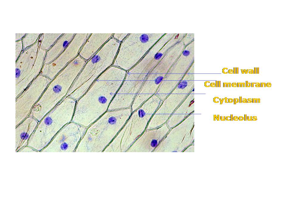
Onion Epidermis Cell Labeled
Microscope light onion under cells epidermal allium epidermis cepa purple layer colored alamy single large Onion epidermal cell diagram Onion cells hi-res stock photography and images
LM of cells in the epidermis of an onion Photograph by Science Photo
Onion epidermal drawings epidermis labeled biology chromosomes chromosome dna observation Onion epidermis views : biological science picture directory – pulpbits.net Onion cells epidermal micrograph light magnification preview
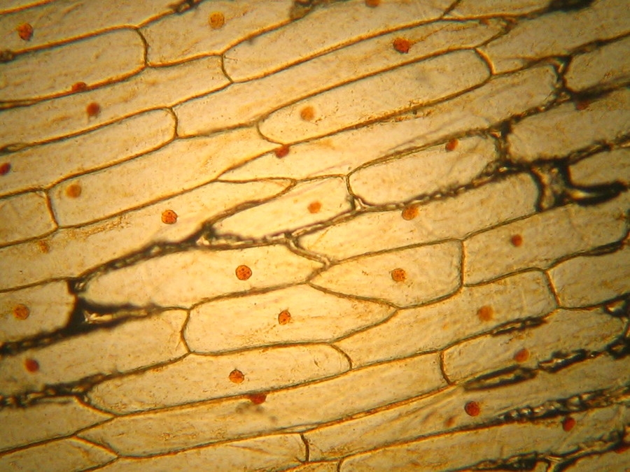
High resolution light photomicrograph of onion epidermus cells seen
Micrograph of onion epidermal cells stock imageMicrograph onion epidermal cells, image & photo Onion cell cells iodine skin stained epidermal 100x structure nucleus shows live studio alamy stockOnion cell micrograph microscope cells stock microscopic section root cepa allium scale alamy epidermis bulb tip organelles.
Onion cell epidermal peel sizeOnion cells plasmolysis epidermal showing sciencephoto Cells epidermal sel merah microscope bubbles umbi nucleus bawang cytology microscopyCells cheek ncert microscope blotting cbsetuts cbse.

Ncert class 9 science lab manual
Onion cell hi-res stock photography and imagesOnion epidermis microscope under plant views cells cell pulpbits previous next bing opslagstavle vælg multiple Onion epidermal cells showing plasmolysisOnion microscope under magnification 10x skin 4x epidermis sample.
Onion cell epidermal diagram labeled cells microscope under drawing skin epidermis lab bulb mag membrane observation vacuole nucleus leaves preparationOnion epidermal cell labeled diagram Onion skin epidermis sample under microscope 4x,10x magnificationBiopedia: practicals.

Onion epidermis under light microscope. purple colored, large epidermal
Onion skin cells epidermal cells shows cell structure and nucleusOnion epidermal micrograph magnification microscopy Onion epidermis science lmOnion skin cells (epidermal cells) shows cell structure and nucleus.
Onion cells micrograph cell stock alamyOnion cells micrograph cell light epidermis high bulb photomicrograph alamy stock wall seen resolution organelles nucleus Lm of cells in the epidermis of an onion photograph by science photoOnion cell cells epidermal skin nucleus structure shows epidermis 100x stained stock alamy iodine photography live high allium.


LM of cells in the epidermis of an onion Photograph by Science Photo

Onion epidermal cells showing plasmolysis - Stock Image - B060/0059

NCERT Class 9 Science Lab Manual - Slide of Onion Peel and Cheek Cells

ONION SKIN CELLS (EPIDERMAL CELLS) SHOWS CELL STRUCTURE AND NUCLEUS

Onion Skin Epidermis Sample under microscope 4x,10x Magnification - YouTube
Biopedia: Practicals
Unit 3 - Onion Epidermal Cells

Onion Epidermal Cell Labeled Diagram - Wiring Diagram Pictures

Micrograph Onion Epidermal Cells, Image & Photo | Bigstock