What Are Onion Cells
Lab slides. cell types Onion cells red file size wikipedia Onion cells slide magnification comments microbiology report
Red onion cells (normal), 100x - a photo on Flickriver
Cells cebola epiderme creativemarket containing micrograph europafotos ukphotos micrografia Onion cells Onion cell
Magnified microscope cell 40x microscopy micrographs walls
Plant cell lab (makeup)Cells, cells, cells – montessori muddle Onion cells 2Image gallery onion cell.
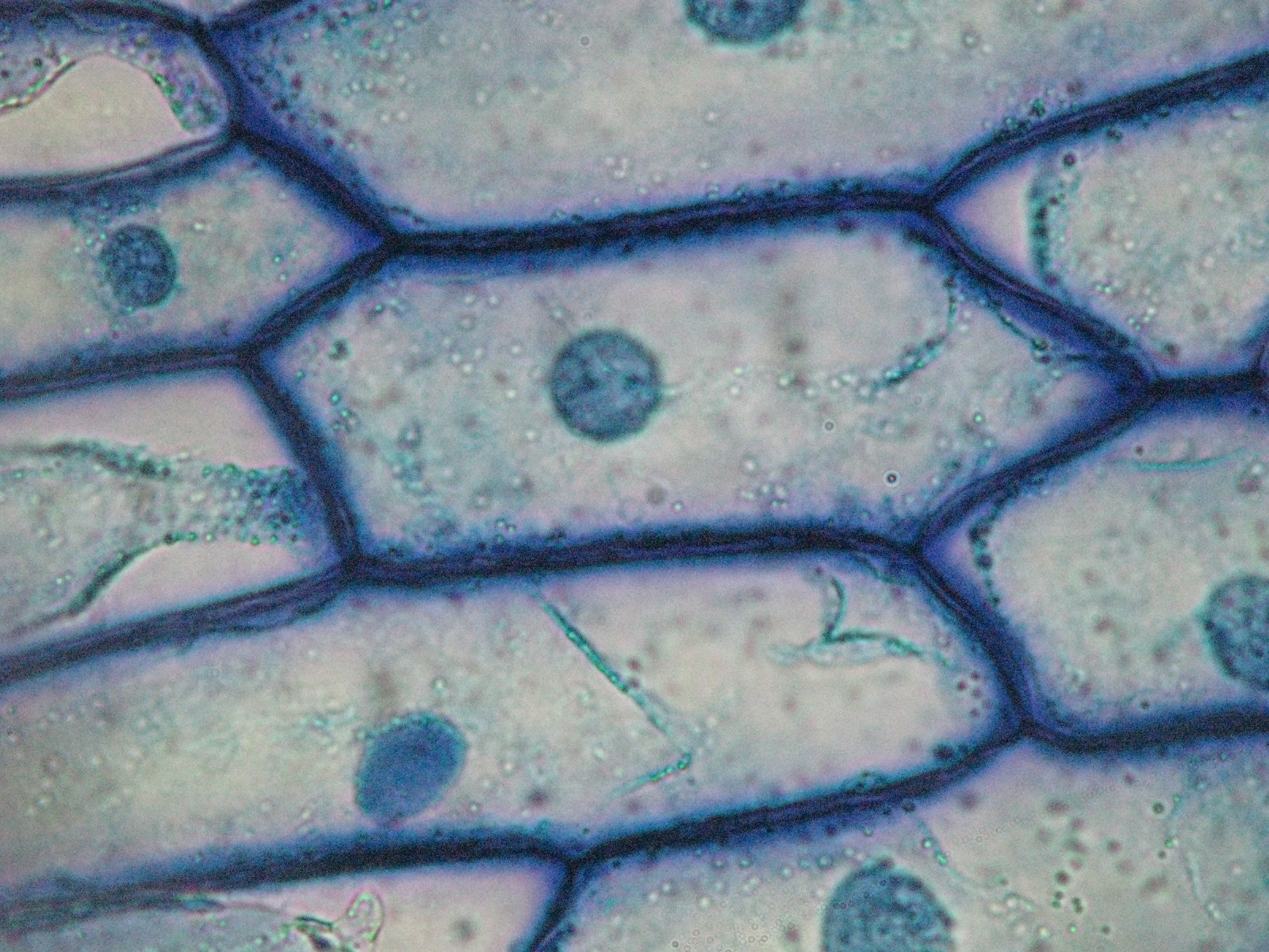
Plant & animal cells staining lab answersCells onion cell microscope under iodine epidermis plant bulb 10x mount inner skin light bubbles air animal nucleus eukaryotic microscopy Onion cell lab red cells stainedMicroscope microscopic staining schoolworkhelper 400x human conclusion.

Onion microscope cell cells under staining lab nucleus nuclei through stained skin slide stain tissue 10x look simple experience dna
Onion cells under microscopeOnion cells Cells onion microscope under skin cell lab 100x lesson fungi algae weebly school plant light magnification differences similarities prokaryotes eukaryotesOnion cell cells microscope micrograph under 40x labeled stock alamy microscopic skin magnification section root tip allium high epidermis bulb.
Onion nucleus microscopesMicroscope electron cell peel diagram badajoz microscopic experiments mitochondria magnification manzano calle estadio vivero microscopy possibilities The inner epidermis of the onion bulb cataphyllsOnion cells cell lab types slides.

Onion cells under microscopes
Onion cells beautiful worldOnion cell microscope hi-res stock photography and images Onion cells microscope under magnified times cell 100x does genetics wallBeautiful world: onion cells.
Onion skin epidermal cells: how to prepare a wet mount microscope slideOnion cells under a microscope Onion cells light micrograph photomicrograph microscope high seen through cell resolution epidermis bulb organelles stock alamy wall scaleSwifty science: onion cell lab.

Cell microscope 40x 400x comparing
Onion cells without microscope staining under observation cell preparation microscopic umberto flickr requirements anyOnion cells slide (80× magnification) : r/microbiology Onion cells stock photos & onion cells stock imagesOnion 40x plant cell lab cells power low biologycorner 400x high 100x makeup biology scanning elodea slide nucleus corner saved.
Image gallery onion cellOnion cells Onion microscope cells skin slide epidermal wet mount plant prepare slides cell animal video stainingOnion cells cell iodine epidermis 10x inner microscopy stained nucleus piece bulb clearly plasmolysis few start show mag.

File:red onion cells.jpg
Onion cells under microscopeRed onion cells (normal), 100x Onion cells cell epidermis bulb tobi looking.
.
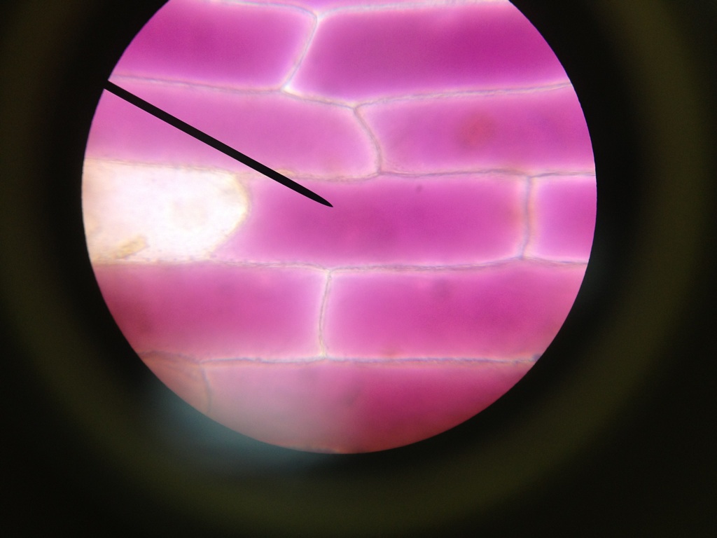

File:Red Onion Cells.JPG - Wikipedia

Onion cells | High-Quality Nature Stock Photos ~ Creative Market

Onion cells 2 | Basically the same of onion cells 1 but this… | Flickr
Beautiful World: Onion cells

Red onion cells (normal), 100x - a photo on Flickriver

Cells, cells, cells – Montessori Muddle

Plant Cell Lab (Makeup)